Tuesday, April 28, 2015

NADPH, the folate cycle, and adrenal function

In my last blog I went over redox balance, the pentose phosphate pathway, and how generation of NADPH by the pentose phosphate cycle impacts adrenal function.  However, recent evidence has identified another metabolic pathway that generates NADPH to provide cellular reducing power, the folate cycle.

NADPH creation by the folate cycle

The folate cycle involves the conversion of dietary folate to different precursors involved in other metabolic pathways depending on cellular needs.


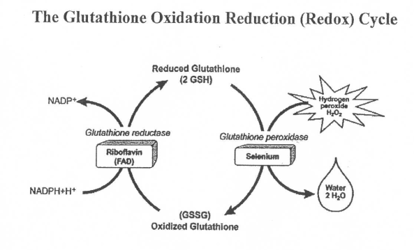
Folate is first converted to THF and then 5,10-MTHF where it enters a crossroads that is partially dictated by the NADP+:NADPH ratio.  NADP+ works with the enzyme MTHFD to direct 5,10-MTHF toward the right in the diagram to help synthesize nucleotides for DNA.  This is the pathway that generates NADPH and inhibition of the MTHFD enzyme increases the ratio of NADP+:NADPH as well as GSSG:GSH(2), shifting redox balance towards oxidation.  As with MTHFR, there are mutations in the MTHFD gene that can negatively impact methylation, likely through changing cellular redox balance.

In the other direction, the enzyme MTHFR converts 5,10-MTHF to 5-MTHF which is used to help power the methylation cycle.  In order for the pathway to operate in this direction, NADPH is necessary as MTHFR is an NADPH dependent enzyme.  This is where it gets interesting.  People with a mutation in the MTHFR gene have lowered activity of the MTHFR enzyme depending on whether they have one or two copies of one of the mutations.  This causes the methylation cycle to function sub-optimally which causes a build up of homocysteine since they don't make sufficient levels of 5-MTHF to help convert homocysteine to methionine.  These people experience reduced enzyme activity merely because they don't produce enough of the enzyme, but that's not the only way MTHFR activity can become reduced.

The activity of the MTHFR enzyme is also dictated by the availability of NADPH, so even people with no MTHFR mutation can experience poor methylation if the redox balance favors NADP+(Oxidation), especially if they have a mutation of the MTHFD gene.  The problem with having reduced MTHFR activity either through a mutation or a less favorable redox balance is that homocysteine may increase free radical production directly(3), by reducing glutathione levels(4), or a combination of the two.  It could also merely be an association where a high level of free radicals is indicative of a cellular environment that will produce more homocysteine, but recent evidence shows that high homocsyteine levels are at least partially causative in increased free radical production(5).  This would promote a more oxidative environment and decrease methylation further.  Maintaining a lower ratio of NADP+:NADPH via the pentose phosphate pathway should help promote a more reductive environment and increase methylation, but people with an MTHFR mutation still likely need to supplement with methylfolate to some extent.

In theory, people with an MTHFR or MTHFD mutation may be more prone to thiamine deficiency than people with the normal SNPs, or at the very least require more thiamine to maintain a more reductive cellular state.  In the MTHFR mutation, if increasing levels of homocysteine change the redox balance to favor oxidative reactions, the pentose phosphate pathway will have to go in to overdrive in an attempt to restore a more reductive cellular environment.  The same could be said with reduced activity of MTHFD since it is an NADPH generating pathway.  Both would require more transketolase activity which will require more thiamine.  Taken together, this could, in theory, mean people with an MTHFR or MTHFD mutation are more prone to adrenal fatigue, assuming the science linking thiamine status to adrenal function is correct.

Blood glucose, insulin resistance, and thiamine

As mentioned in my last blog, thiamine is used in more processes than the pentose phosphate pathway, specifically the pyruvate dehydrogenase and alpha-ketoglutarate dehydrogenase complexes in the mitochondria.  If thiamine is tied up producing reducing equivalents in the pentose phosphate pathway in the cytosol, it may not make it to the mitochondria and could negatively impact the ability to generate energy from glucose there aerobically.  Furthermore, since thiamine is transported in the blood and red blood cells utilize large amounts of thiamine in the pentose phosphate pathway because they are exposed to large numbers of free radicals, cells that utilize glucose may not get the thiamine they need to metabolize glucose for energy effectively when free radical levels are high, as in hyperglycemia.  Decreased thiamine in cells that rely on oxidative glucose metabolism will decrease pyruvate dehydrogenase activity, increasing lactate production.  This would increase blood lactate levels which is known to induce insulin resistance in skeletal muscle(6), forming a vicious cycle where free radical production induced by hyperglycemia increases insulin resistance and reinforces hyperglycemia, leading to more free radical production.

This is not the only problem facing people with Type 2 diabetes.  In an ironic twist of fate, Type 2 diabetics have altered thiamine metabolism.  One study found marginally low plasma thiamine levels in people with Type 2 diabetes that translated in to reduced transketolase activity(7).   Another study found plasma thiamine levels in people with Type 2 diabetes to be reduced by 75% compared to controls with a 16x increase in renal thiamine excretion(8).  A yet to be published observational study found between 16%-29% of obese people seeking bariatric surgery to be deficient in thiamine(9) and a study in Australia found low plasma thiamine and folate in healthy blood donors(10), so this may not simply be an obesity/Type 2 diabetes problem but a problem of the modern diet.  While the US healthcare system views thiamine deficiency as being relatively rare and only in alcoholics, it appears the data doesn't really support this position.

One of the more beneficial aspects of undertaking a Paleo diet is that it can have a profound effect on reversing Type 2 diabetes(11, 12).  However, based on the available evidence, one may need to move from their old lifestyle to their new one with caution.  If a person with Type 2 diabetes decides to move from the Standard American Diet to a Paleo diet and their inability to properly regulate blood glucose during their old diet put them at marginal or low thaimine status, cutting out grains and legumes, two significant sources of thiamine, may sink them deeper in to thiamine deficiency.  Throw in a few days of glucose demanding Crossfit WODs per week and the stage for thiamine deficiency, and perhaps adrenal fatigue, is set.  Supplementing with thiamine or making sure the diet provides adequate thiamine is the prudent course for those switching from a standard American diet to the Paleo diet.

Conclusion

At this point, I assume your head is spinning so let me break the last 2 blogs down for you.  Cellular redox balance is important to cellular function as it helps dictate the direction of metabolic pathways. Two redox pairs exist in different ratios to allow anabolic and catabolic reactions to go on, sometimes at the same time, in a cell.  The acronym ARCO can help you remember that anabolic reactions tend to favor reduction while catabolic reactions tend to favor oxidation.  The low ratio of NADP+/NADPH favors reduction which makes it an anabolic coenzyme pair, but this balance can be thrown off if cellular free radicals rise and glutathione uses the reduction power of NADPH generated from the pentose phosphate pathway to maintain its ability to neutralize these free radicals. Low thiamine intake may also throw off cellular redox balance by reducing flux through the pentose phosphate pathway, reducing NADPH levels.

Looking at the folate and methylation cycles, reduced activity of the MTHFR enzyme, which is dependent on NADPH, will increase free radicals through increased homocysteine production.  Again, this would drive the redox balance towards oxidation via increased glutathione reduction. MTHFD, the enzyme responsible for directing the folate cycle in the opposite direction, is also of concern because it contributes significantly to the pool of NADPH in the cell. People with mutations in the genes that code for these enzymes likely have increased thiamine needs, particularly if their lifestyle leads to high levels of free radicals due to blood glucose fluctuations, high alcohol intake, or smoking.

Now, with the hard science over, in the next blog we can go over a more easy to understand mechanism by which thiamine deficiency can impact adrenal function: altered synthesis of the neurotransmitter acetylcholine. 

Tuesday, April 21, 2015

Redox balance, the pentose phosphate pathway, and adrenal function

In my last blog I went over some of the science linking thiamine deficiency to altered adrenal function, dysautonomia, and how that relates to adrenal fatigue and it's symptoms.  In this blog we begin looking at mechanisms by which thiamine impacts adrenal function.  The first mechanism deals with the pentose phosphate pathway as well as the folate cycle.

Cellular redox balance

Before we get in to the specifics of how the pentose phosphate pathway and folate/methylation cycles affect adrenal function, we need to discuss something called redox balance.  Redox reactions involve the passing of electrons between molecules and are normally coupled with one another.  Reduction involves a molecule gaining an electron while oxidation involves a molecule losing an electron.  In order for one molecule to gain an electron, one must give up an electron, hence the pairing. 

You may be familiar with free radicals and antioxidants.  Free radicals are molecules that have an unpaired electron in their outer shell.  This makes them unstable so they "steal" electrons from other molecules.  By stealing an electron, a free radical becomes more stable and is reduced while the other molecule becomes unstable and is oxidized.  Antioxidants donate an electron to free radicals to prevent healthy tissues from becoming oxidized, but when they reduce free radicals they become oxidized and unstable themselves.
 
Based on the above information, we can call free radicals oxidizing agents and antioxidants reducing agents.  Redox balance refers to the reactive state of the cell.  A cell with a higher percentage of oxidizing agents will favor oxidation while a cell with a higher percentage of reducing agents will favor reduction.   In addition, certain redox pairs exist in different ratios since they function as coenzymes in metabolic pathways.  This is important because many biochemical reactions are dependent on cellular redox balance and this balance will dictate the direction of the pathway as each side of the coenzyme pair causes the reaction to go in a different direction.

Think of it this way.  Often times, when a molecule comes to a metabolic crossroads, it encounters 2 enzymes that will direct it in opposing directions.  Each one of these enzymes is dependent on a cofactor for activation.  If the reduced cofactor is present in higher concentrations, the enzyme dependent on the reduced coenzyme will become active while the one dependent on the oxidized cofactor will be more dormant.  This will direct the molecule down that enzymes pathway and oxidize the cofactor, increasing the chances that the next one of those molecules will go in the other direction.  However, these cofactors are used in so many different reactions that it's possible to "lock" the cellular pathway to favor oxidation or reduction if the redox balance favors one or the other.

The three primary coenzyme redox pairs are FAD/FADH2, NAD+/NADH and NADP+/NADPH; noted as oxidizing agent/reducing agent.  Since cells tend to maintain a very high ratio of NAD+:NADH(Approximately 700 in mammalian tissues), this coezyme pair favors oxidation while the very low NADP+:NADPH ratio in cells(.005) favors reduction.  This allows cells to perform both oxidation and reduction depending on whether the enzyme in the reaction prefers NAD+/NADH as the coenzyme pair or NADP+/NADPH.  In addition, some of these pairs work together as coenzymes, passing electrons between one another.  FAD/FADH2 often work in concert with NADP+/NADPH as cofactors for certain enzymes, many of which are involved in adrenal function. To keep it simple, for the purposes of this blog, we will focus on NADP+/NADPH.  Keep in mind, as mentioned above, that once NADPH is used in a reaction it becomes NADP+, and vice versa.  We use the term redox balance because when one side of the pair goes down the other goes up.

NADPH and cellular redox balance

NADPH is a very interesting molecule.  It's used in cells to provide reducing power to promote anabolic reactions as well as function as an electron donor to glutathione.  Glutathione functions as the primary cellular antioxidant and exists in a reduced (GSH) and oxidized (GSSG) form.  When GSH encounters a free radical, it donates an electron with the help of selenium to stabilize the free radical and becomes GSSG, its oxidized, inactive form.  NADPH, in concert with riboflavin(FAD), then converts GSSG back in to the active GSH.  This process converts NADPH to NADP+.
This cycle occurs over and over again in your cells as they encounter free radicals.  Therefore, a high level of free radicals in the cell will shift the redox balance towards oxidation.  However, as you may notice on the left side of the diagram, we have yet to discuss how NADP+ gets converted back to NADPH so that it can reactivate GSSG to GSH again and promote a more reductive cellular environment.  This is where the pentose phosphate pathway comes in.  The oxidative phase of the pentose phosphate pathway converts NADP+ to NADPH to help maintain a reductive state(More NADPH in relation to NADP+).

The non-oxidative phase supports this process by converting products of the oxidative phase back in to glucose 6-phosphate to create more NADPH via the enzymes transaldolase and thiamine dependent transketolase.  For every molecule of glucose 6-phosphate, the pentose phosphate pathway can create 2 NADPH from NADP+ using only the oxidative phase while using both phases yields 12 NADPH provided there is enough thiamine to maintain transketolase activity.  Keep in mind, when looking at redox balance, this means that the oxidative phase increases the number of NADPH by 2 and also decreases the number of NADP+ by 2 while the non-oxidative branch changes each by 12, a 24 point swing in redox balance in favor of NADPH.

Redox balance, specifically NADP+:NADPH, relates to adrenal function because biosynthesis of glucocorticoids, as well as most steroid hormonse, is dependent on NADPH(1).  This could help explain why thiamine deficiency has such an impact on adrenal function because thiamine, specifically thiamine diphosphate, is necessary to get the full NADPH recharging effect of the pentose phosphate pathway.  Additionally, NADP+ favors the conversion of cortisol to cortisone, a weaker glucorticoid, while NADPH favors the opposite conversion.  A redox balance that favors oxidation in the adrenal glands, therefore, can have a negative impact on adrenal function by creating more cortisone than cortisol.  It's interesting to note that cortisol is also capable of binding to the mineralocorticoid receptor while cortisone is not.  This would negatively impact electrolyte balance by increasing sodium loss in the urine, a common casuative factor in adrenal fatigue symptoms.

While we have focused on the pentose phosphate pathway for NADPH production because it provides the greatest contribution, there are other ways NADPH can be produced.  One newly discovered and very interesting pathway involves the folate cycle, so if you have an MTHFR mutation, you may want to strap in.

Next: NADPH, the folate cycle, and adrenal function

Tuesday, April 14, 2015

The importance of addressing thiamine status in adrenal fatigue

In my last blog I discussed the multi-system symptomology of adrenal fatigue and used the analogy of a home heating system to describe how one may address the underlying causes of adrenal fatigue.  The analogy identified 3 components of your home heating system that may be the problem.
  1. The thermostat isn't set properly or doesn't sense the temperature
  2. The ignitor doesn't turn the gas in to heat
  3. The gas flow is off or obstructed
In this analogy, properly setting the thermostat involves changing your lifestyle to address how your brain perceives stress while fixing gas flow is increasing carbohydrate or caloric intake.  Both of these components are important factors to consider and most people do a good job at addressing them.  Addressing the ignitor, on the other hand, is another story.  I would consider addressing the ignitor as addressing nutritional deficiencies.  One nutritional deficiency that has some pretty solid science behind it is thiamine deficiency.  Most people are quick to address vitamin C, magnesium, D3 and other deficiencies with large doses of vitamins or multivitamins while ignoring something that may be as, if not more, important.  Let's take a look how thiamine may play a role in adrenal fatigue.

Thiamine 101

Every living organism on the planet, from bacteria to plants to animals, requires thiamine.  Certain bacteria and plants can synthesize thiamine on their own but animals require thiamine in their diet.  Thiamine is found in a variety of foods including yeast, lean pork, grains, legumes, and certain seeds.  Liver also contains a large amount of thiamine as most animals, including humans, have high stores of thiamine in the liver and red blood cells.

Thiamine is absorbed from the jejunum and ileum from food that is digested or, in some cases, via production by resident gut bacteria.  In fact, of the 3 identified human enterotypes, enterotype 2 has a large proportion of thiamine generating bacteria making hosts with that enterotype less likely to experience thiamine deficiency(1).  There are also bacteria that bind thiamine or create thiaminases, enzymes that degrade thiamine.  Humans absorb a high percentage of low dose thiamine but a gradual decline in the percentage of thiamine absorbed occurs at levels above 5 mg.  Thiamine is absorbed by intestinal cells as thiamine diphosphate but is converted in to free thiamine and released in to the bloodstream.    In the blood, it circulates as free thiamine and only becomes active when it is phosphorylated.  The most active form of thiamine is thiamine diphopshate although it seems thiamine triphosphate has some important, not well defined roles in the nervous system.

Humans store between 25-30mg of thaimine, much less than other animals.  Due to thiamine being a water soluble nutrient, depletion can occur in 14-18 days.  Under deficient thiamine intake, different organs lose thiamine at different rates.  Of utmost importance, the brain and central nervous system hold on to thiamine much longer than other organs.  This is likely due to the brains reliance on oxidative glucose metabolism and the role thiamine dependent enzymes play in that process.  The limbic system, an area of the brain responsible for emotion that also contains the hypothalamus, is typically hit very hard by thiamine deficiency.  It's of interest to note for our purposes that the hypothalamus is the H in the HPA axis. 

Cellular roles of thiamine

Thiamine has several roles in cellular glucose metabolism as it functions as a cofactor for various enzyme complexes.  The pyruvate dehydrogenase(PDH) and alpha ketoglutarate dehydrogenase(a-KGDH) enzyme complexes are important thiamine dependent enzyme complexes that help liberate energy from glucose in the citric acid cycle of mitochondria.  During glycolysis in the cytosol, glucose is converted in to 2 pyruvate molecules that enter the mitochondria.  Inside the mitochondria, pyruvate is converted in to acetyl CoA by the PDH complex so that it can enter the citric acid cycle.  This step requires thiamine diphosphate as a coenzyme.  This is important for 2 reasons.  In neurons, acetyl CoA comes predominantly from glucose and is necessary for the synthesis of the neurotransmitter acetylcholine, which we will cover later.  Secondly, in all cell types, insufficient thiamine decreases PDH activity and lactate accumulates in the cell and pours out in to the circulation.  Blood lactate is known to be elevated in Type 2 diabetics(2) and high blood lactate levels induce insulin resistance in skeletal muscle(3).

The role of a-KGDH in the citric acid cycle is also of importance as this enzyme complex is necessary for the synthesis of the neurotransmitters GABA, glutamate, and aspartate.  Furthermore, the altered glucose metabolism that accompanies a deficiency in the activity of PDH and a-KGDH can lead to mitochondrial damage and eventual cell death(4).

Another area of glucose metabolism where thiamine is important is the pentose phosphate pathway.  The pentose phopshate pathway is an anabolic pathway of glucose metabolism that creates NADPH or R5P based on cellular needs.  For a more thorough look at this process, check out this blog.  Understanding the pentose phosphate pathway is crucial for understanding hormonal balance and how adrenal function can be affected by thiamine deficiency so I urge you to check that blog out.

The thiamine dependent enzyme in the pentose phosphate pathway that's important is called transketolase.  Transketolase allows the products of  the non-oxidative pathway of the pentose phosphate pathway to be recycled in to glycolysis for generation of energy, to be converted in to glucose 6 phosphate to re-enter the oxidative phase of the pentose phosphate pathway to generate NADPH, or it can work in reverse and convert glycolytic intermediates in to ribose 5 phosphate, a necessary component of DNA and RNA.

This diagram shows the function of transketolase, abbreviated as Tkt.  Note how transketolase allows the products of the pentose phosphate pathway to move back in to glycolysis or feed back in to the pentose phosphate pathway.  One could look at it as transketolase preventing metabolic dead ends in the pentose phosphate pathway that aren't really dead ends at all.  Without transketolase, these products may accumulate and enter pathways that lead to glyoxal and methylglyoxal formation that eventually lead to advanced glycations endproducts(AGEs).  Thiamine has been shown to decrease formation of these troublesome substrates(5, 6) and the primary mechanism is through increased transketolase activity(5, 7) re-routing precursors back in to the pentose phosphate pathway and away from glyoxal formation.
 

Research on thiamine deficiency and adrenal function

Given the ethical challenges that inducing a thiamine deficiency in humans would raise, much of the data on the effect of thiamine deficiency on adrenal function comes from studies in rats.  One study showed that inducing thiamine deficiency in rats led to hyperstimulation of the zona fasciculata of the adrenal glands in 2 weeks causing increased corticosterone output followed by complete exhaustion in 4 weeks(8). Corticosterone is the chief glucocorticoid in rats whereas cortisol fills that role in humans.  While this is obviously an extreme example of thiamine deficiency and its effect on the adrenal gland, it does underscore the importance of thiamine in adrenal function.

Another study in rats found thiamine deficiency elevated corticosterone levels and depressed the aldosterone response to sodium deprivation(9).  Aldosterone is released by the adrenal glands when sodium levels drop, causing the kidney to recycle sodium back in to the bloodstream.  This is interesting because many of the symptoms associated with adrenal fatigue relate to an electrolyte imbalance, specifically a decrease in the sodium:potassium ratio.  A decrease in aldosterone under low sodium intake would induce the same set of symptoms.  Many people with adrenal fatigue notice an improvement in their symptoms with increased salt intake.

A study in humans found thiamine injections prevented functional adrenal gland exhaustion during and after surgical stress(10).  Again, it's hard to extraploate this data to otherwise healthy individuals, but it does show a general effect of thiamine on adrenal gland function.  Other studies in humans, particularly alcoholics, show biochemical lesions in the brain of people who are thiamine deficient.  This is likely due to decreased a-KGDH activity and impaired carbohydrate metabolism(11).  Since these lesions manifest themselves in the limbic system of the brain, they likely have an effect on adrenal function via an altered emotional state as well as damage to the hypothalamus.

It is apparent that thiamine is important for proper adrenal function.  The question now becomes what are the mechanisms by which thiamine deficiency can lead to adrenal dysfunction.  We'll tackle that after the break.

Redox balance, the pentose phosphate pathway, and adrenal function

Thursday, April 2, 2015

The multi-system symptomology of adrenal fatigue: Is thiamine deficiency at play?


People with adrenal fatigue tend to have symptomology that ranges across many body systems.  While these systems likely affect one another due to the fact that they must work in concert with one another to help us adapt to our environment and are controlled by the autonomic nervous system, it's an assumption that one system is throwing the others out of whack.  While this may be true, there is the potential that what we are seeing in adrenal fatigue isn't just one system throwing other systems off, but all systems being thrown off by a deficiency in a nutrient that they all rely on for proper function. 

Dr. Derrick Lonsdale, MD has written many articles on dysautonomia, dysfunction of the autonomic nervous system, which is the defining characteristic of adrenal fatigue.  He points to dysfunction in oxidative carbohydrate metabolism as the primary cause of dysautonomia(1).  He discusses the early stages of beriberi, a disease of thiamine deficiency, as the prototypical example of dysautonomia(2, 3).  His perspective is coming from the Standard America Diet and it's reliance on processed carbohydrate as being causative in thiamine deficiency.

It is interesting to note that beriberi was discovered as being caused by an imbalance between the level of dietary carbohydrate and thiamine.  In 19th century Japan, beriberi was extremely common in the Japanese Navy and the culprit was eventually determined to be diet related.  Rice that has been polished, white rice, is stripped of its thiamine content while leaving the carbohydrate levels intact.  Cadets who had relied solely on white rice were far more likely to experience beriberi than cadets fed a more varied diet.  This led to the discovery of accessory nutrients, aka vitamins, that were necessary for proper cellular metabolism.

It is assumed in modern medicine that the only people who experience thiamine deficiency are alcoholics or the malnourished.  Dr. Lonsdale and his co-workers have published multiple case studies showing thiamine deficiency as a product of micronutrient deficiency brought on by excess processed carbohydrate consumption.  Dr. Lonsdale calls this high calorie malnutrition.  These people are neither alcoholics nor malnourished by macronutrient standards.  Many of these people are told that their symptomns are in their head by their mainstream doctor, and when they are tested for thiamine deficiency by Dr. Lonsdale they are shown to be deficient because a mainstream doctor isn't on the look out for thiamine deficiency.  Thiamine defiency is known to affect the limbic system very hard.  The limbic system is an area of the brain responsible for emotion, adrenaline flow, motivation, long-term memory, and contains the hypothalamus: the H in the HPA axis,.

The symptomology of these case studies closely reflects autonomic dysfunction, similar to the early stages of beriberi(3), and are corrected by increased thiamine intake.  As mentioned above, thiamine needs are known to be dependent on carbohydrate intake, but the question is are they dependent simply on carbohydrate intake or are they also dependent on how much a person relies on oxidative carbohydrate metabolism for their physical activity?

In people on the Standard American Diet, high intake of processed carbohydrate in the absence of adequate thiamine presents as a thiamine deficiency because they are forcing glucose in to their cells which increases their need for the nutrients needed to efficiently oxidize glucose.  While these people may meet the RDA for thiamine, these RDAs were likely determined based on a lower consumption of carbohydrate.  Eating larger doses of carbohydrate with the same level of thiamine may actually reflect deficiency as cells are unable to oxidize the level of carbohydrate contained in the diet.  In addition, higher levels of free radicals brought on by hyperglycemia may require higher thiamine intake to produce NADPH in the pentose phosphate pathway for reduction of these free radicals via reduction of glutathione(We will cover this in the next blog).  Another problem is that people with insulin resistance and type 2 diabetes have dysregulated thiamine status evidenced by a 75% reduction in plasma thiamine levels in comparison to controls(4), most likely due to thiamine loss in the urine.  It's also interesting to note that these people also tend to be sedentary, so muscle stores of thiamine are likely to be fairly low as well.

Participating in intense exercise that relies on these same glycolytic pathways should cause the same problem.  Compounding the issue is that people doing this who also eschew grains and legumes are eliminating 2 of the better sources of thiamine in the diet.  One could probably meet thiamine needs with other food sources, particularly liver, the question is are you?  If a person is already at marginal thiamine status from insulin resistance or random bouts of hyperglycemia and they cut out 2 significant sources of thiamine, deficiency seems likely.  One has to question what would happen if a person with Type 2 diabetes/insulin resistance went from eating the Standard American Diet with already low to marginal thiamine status to cutting out grains and legumes from their diet and exercising intensely.  Sounds like a recipe for autonomic dysfunction, aka adrenal fatigue.

Hopefully this blog has put thaimine on your radar screen, particularly if you plan to undertake a diet such as the Paleo diet, which I hope you do.  A nutrient dense diet that limits processed food is universally considered the optimal human diet.  However, one has to be sure to meet thiamine requirements as well as not overdo the intense exercise portion of the lifestyle right off the bat.  Before you go out to the store and buy regular old thiamine, let me save you the time, if you already have adrenal fatigue it's not going to work.  Don't worry, we'll get to that later.  In the next blog we will look at the science of how thiamine deficiency affects adrenal function.

The importance of addressing thiamine status in adrenal fatigue