Thursday, October 29, 2015

Methylation and the folate cycle: You're on the wrong path(way)!

File:Choline metabolism-en.svg 

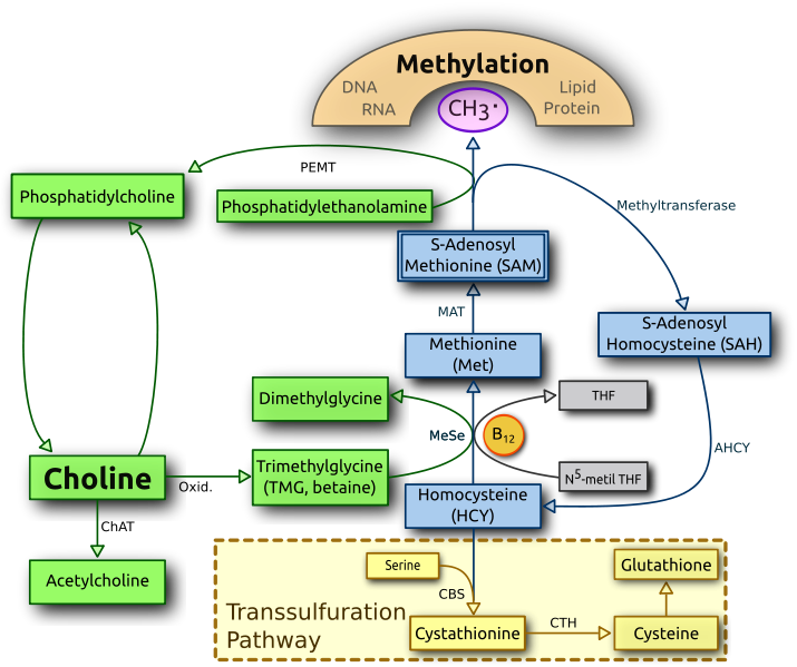
The picture above represents the methylation cycle and the pathways that help power it.  On the right side in grey you have a partial representation of the folate cycle while on the left side we have a mostly complete representation of choline metabolism.  Both of these processes intersect the methylation cycle at the same point, a critical point where homocysteine is recycled in to methionine.  High homocysteine levels are a bad scene as homocysteine is toxic, so having efficient conversion of homocysteine to methionine is important to prevent this scenario.

The folate cycle and choline metabolism balance each other out

Many people are aware of how the folate cycle impacts methylation, many are even aware of gene polymorphisms they have that negatively impact this point of the methylation cycle.  However, few realize that having a defect on one side is typically compensated for by the other side.  In other words, when the folate cycle doesn't run well, choline metabolism can pick up the slack with respect to the methylation cycle and convert homocysteine to methionine, and there are studies that show this(1, 2).  It's important to understand the opposite to be true as well, when choline metabolism doesn't work well the folate cycle can pick up the slack with regard to the methylation cycle.

The problem is that most people with polymorphisms related to the folate cycle focus on getting more folate or increasing methyl-B12 in the hopes of improving methylation.  This is a fool's errand as the primary polymorphism occurs in the MTHFR gene, which is the rate limiting step and occurs prior to the step in the cycle where homocysteine is recycled to methionine.  This isn't to say that you shouldn't get adequate B12 and folate, just that this pathway will always be inefficient at recycling homocysteine to methionine.  Why focus on it then?

Dietary choline vs. choline synthesis

点击查看完整图片

Looking at the choline side of the equation we have 2 pathways to ensure adequate choline, dietary intake and synthesis from phosphatidylethanolamine(PE).  As you can see from the figure, in order for phosphatidylethanolamine to become phosphatidylcholine(PC) which can then become choline, you have to use SAM-e which then becomes homocysteine.  To be precise, you use 3 SAM-e so you make 3 homocysteine molecules.  This pathway is obviously not ideal if you wish to lower homocysteine levels as it also increases them, so adequate dietary intake of choline is the optimal pathway.  In someone with polymorphisms in the genes that negatively affect the folate cycle, choline intake is very important as a poorly functioning folate cycle increases choline requirements(1, 2).  Ingesting choline in the diet decreases homocysteine because it provides an eventual methyl group to convert homocysteine to methionine AND it prevents the generation of homocysteine through choline synthesis from phosphatidylethanolamine.

Choline is not considered an essential nutrient in the United States by definition because people who get adequate methionine can make it and people with adequate folate intake need less of it.  The problem with this definition is that inadequate choline increases the demand for both methionine and folate and it does not take into consideration polymorphisms, such as MTHFR, that increase choline requirement.  This caused the Institute of Medicine to reclassify choline as an essential nutrient. 

The recommended intake for choline is set at 425mg/day for women and 550mg/day for men, but people vary in their need for choline.  People with MTHFR polymorphisms as well as pregnant women need higher intakes of choline.  Foods that are high in choline include eggs, oysters, fish, poultry, beef liver, cruciferous vegetables and peanuts.  Supplementing choline in the form of soy or egg lectithin, choline bitartrate, or phosphatidylcholine is also effective and well tolerated.

Signs you need more choline

One of the easiest signs that you need more choline is that you are a human, living in the United States.  Only 10% of older children and adults get adequate choline in their diet and not all show overt clinical symptoms(3).  Fatty liver and muscle damage are common, but these symptoms tend to be subclinical in nature so most people would have no idea they are experiencing this until something significant happens.  I would suspect that most symptoms of choline deficiency would primarily center around functions that rely on methylation or the neurotransmitter acetylcholine.  Since most people who are currently trying to improve the function of the folate cycle are familiar with symptoms associated with poor methylation, I'll focus on the symptoms associated with low acetylcholine.

Acetylcholine is formed when the enzyme choline acetyltransferase turns acetyl CoA and choline in to acetylcholine and coenzyme A.


Many executive functions such as sleep, stress, digestion, GI motility, and memory all rely on sufficient acetylcholine.  In regard to sleep, acetylcholine is very important for REM sleep, the stage of sleep where dreams, and potentially memory consolidation, occur.  Within the autonomic nervous system, preganglionic nerves of both the sympathetic and parasympathetic branches use acetylcholine as well as postganglionic nerves of the parasympathetic branch.  Symptoms of acetylcholine deficiency in the autonomic nervous system would likely include anxiety and mood disorders.

During digestion, acetylcholine is responsible for the secretion of enzymes in the stomach as well as motility throughout the digestive tract.  Impairments in these functions can lead to IBS, SIBO, or poor nutrient status due to problems in absorption.  An interesting side note is that both caffeine and nicotine, which tend to increase GI motility, work by increasing acetylcholine levels or activating acetylcholine receptors.  Caffeine works by inhibiting the breakdown of acetylcholine and nicotine works as a substitute for acetylcholine by stimulating nicotinic acetylcholine receptors.  Linking digestion and the autonomic nervous system together, the primary neurotransmitter used by the vagus nerve is acetylcholine.  The vagus nerve is known as the conduit through which the brain and gut communicate with one another.

Finally, with regard to memory, acetylcholine is very important for the formation of new memories and deficiency can impair working memory(4).  This would most likely present as brain fog.  Further underscoring the importance of acetylcholine for memory, the class of drugs most used for Alzheimer's disease work by inhibiting the same enzyme that coffee inhibits and people with Alzheimer's disease produce less acetylcholine and have dysfunctional nicotinic acetylcholine receptors(5).

So, it's as simple as supplementing with choline, right?  If only it were that simple...

Monday, October 12, 2015

The migrating motor complex: Housekeeping system for the digestive tract

Have you ever felt a rumble in your tummy when it's been a while since you've eaten anything?  Do you know what it means?  While most people think it's a signal that they need to eat, it's actually the migrating motor complex, the mode your digestive system goes in to when you are in a fasted state.  The purpose of the migrating motor complex isn't to inform you that you are hungry, that's what your appetite is for.


The migrating motor complex

The migrating motor complex is actually housekeeping mode for your digestive tract.  The basics of it are simple.  Throughout your digestive tract, particularly the stomach and duodenum, there are receptors that detect nutrients.  When these receptors no longer detect nutrients and pressure in the duodenum increases through light contractions of the smooth muscle, the migrating motor complex is initiated.  Serotonin builds up in the duodenum and causes secretion of the hormone motilin.  Motilin causes strong contractions starting in the stomach that work their way down the digestive tract to the end of the small intestine, called the ileum. If you're interested in the hard, dirty science of it, the schematic below should help:


These rhythmic contractions of the digestive tract occur during interdigestive periods to move undigestible material, leftover particles of food, and unwanted bacteria from the stomach and small intestine in to the colon where they can be eliminated in feces, a process known as peristalsis.  During this process, there is also an increase in enzymatic secretions that aid in cleansing the digestive tract by breaking down undigested food particles and killing bacteria.  It is interesting to note that people with small intestinal bacterial overgrowth(SIBO) have a defective migrating motor complex.  Unfortunately, this is a classic chicken or the egg scenario so we don't know if it's cause or effect, but it's likely both.

There are 4 phases in the migrating motor complex, phase 3 being the most important as it is the period of greatest motor and secretory activity.  When you feel that rumbling in your belly, those are phase 3 contractions of the stomach.  Phase 3 lasts between 5-15 minutes and occurs roughly every 90-120 minutes.  Since this is the migrating motor complex, and the end goal is to move contents of the digestive tract to the end for eventual elimination, phase 3 contractions do not all happen at the same time.  The contractions typically begin in the stomach and move progressively through the beginning(duodenum), middle(jejunum), and end(ilieum) of the small intestine.  Sometimes the phase 3 contractions can bypass the stomach and begin in the duodenum or jejunum, but the vast majority begin in the stomach.


Factors that affect the migrating motor complex

Multiple factors have an impact on the migrating motor complex.  The primary factor regulating it is the presence of food in the digestive tract.  Once food is consumed, phase 3 contractions cease no matter which part of the digestive tract they are happening in.  This is because the types of muscular contractions that occur during digestion are distinct from those of the migrating motor complex.  While the contractions of the migrating motor complex are meant to clean and move food out of the digestive tract, the contractions that occur during digestion are meant to mix and grind the food you eat, which occurs in a distinctly different fashion.

Another factor is stress.  The digestive tract contains its own, fully functioning nervous system called the enteric nervous system.  The enteric nervous system can function on its own without input from the central nervous system, but the central nervous system does interface with the enteric nervous system and imparts some control over it through the autonomic nervous system, your stress management headquarters.  Specifically, the vagus nerve and prevertebral ganglia interface with the enteric nervous system.  While this control is not necessary, it is certainly optimal as the vagus nerve controls acid secretion and motility of the stomach.  Specifically with regard to the migrating motor complex, stress delays phase 3 contractions in the stomach and likely impacts gastric acid and pepsin secretion.

The final factor we will discuss that impacts the migrating motor complex is sleep.  Sleep slows down the migrating motor complex.  This is unfortunate because the vast majority of people spend most, if not all, of their time fasting asleep. This means that, in most people, digestive housekeeping more like Animal House and less like Grandma's house.


Modern eating patterns are no good for a clean GI tract

If you think about it, the eating patterns of most people probably doesn't lend itself well to this entire housekeeping process, including those that are living a "healthy" lifestyle.  If you eat 5 times a day with the first meal being as soon as you wake up, exactly when do these interdigestive periods begin?  It takes approximately 2-6 hours for food to leave the stomach and an additional 3-5 hours for food to leave the small intestine.  Given the fact that each migrating motor complex takes, at minimum, 90 minutes to complete, you'll likely get no full cycles during the day.  This means you are leaving the entirety of the housekeeping process to the times when it is least active, at night.

In my opinion, 5 meals a day really serves no purpose.  For one, the thermic effect of food, the energy demanding process that would be impacted by what you eat, isn't even dictated by how often you eat, it's dictated by how much you eat.  Those same receptors in your stomach that detect nutrients use that information to determine how much digestion needs to take place, how many types you eat doesn't even factor in to the equation.  Even eating 3 times a day may be too much because there really isn't an interdigestive period during the day.  In certain circumstances it may be necessary to ingest smaller meals, particularly if you have poor digestion.  However, this may not be optimal and a better approach may be to ingest digestive enzymes with your food until your digestion gets back on track.

Probably the most effective way to get all you can out of this housekeeping process without drastically changing your lifestyle is to withhold ingesting anything with calories until at least 3 hours after you wake up.  Notice I didn't say eating, I said ingesting anything with calories.  You can drink coffee, just stop dumping butter or coconut oil in it.  Black coffee has a stimulative effect on peristalsis and may increase gastric secretions because caffeine blocks the breakdown of acetylcholine.  This would, in theory, improve the effectiveness of the migrating motor complex by strengthening contractions and enzymatic secretions.  To fully optimize the process, withhold eating anything at least 3 hours before you go to bed.

How can I tell if it's working?

A great way to see if your migrating motor complex is working is to pay attention to what your stomach is telling you.  That rumbling in your belly is a good thing.  If you aren't getting that rumbling, something isn't working properly.  There is really no way to know for sure, but, in my opinion this whole process is likely controlled by bacteria in your gut.  The synthesis and secretion of serotonin and acetylcholine, two neurotransmitters that are used heavily in the migrating motor complex, seem to be modified by the bacteria in your gut.  If this is the case, what you eat is probably as important as how often you eat for efficient housekeeping of the digestive tract, something we may cover in a future blog.

Tuesday, July 21, 2015

Chronic Traumatic Encephalopathy: Are there ways to mitigate risk?

In recent years, research in to Chronic Traumatic Encephelopathy(CTE) has exploded as the number of professional football players and war veterans who have committed suicide has increased.  The chronic condition, thought to be a consequence of repeated blows to the head in sports such as football and boxing or from blast-exposed military veterans, leads to progressive loss of brain tissue and abnormal brain physiology somewhat similar to that of Alzheimer's disease patients.  The disease is insidious, beginning with mood disturbances and psychotic episodes early on.  In the second stage, memory loss, erratic behavior, and Parkinson's symptoms become increasingly prominent.  Finally, victims of the disease experience dementia, full-blown Parkinson's, and speech and motor dysfunction.  While progression of the disease is often blamed solely on repeated blows to the head sustained as a result of contact sports, oftentimes the symptoms don't appear until a much later time, sometimes a decade or more after retirement.  Making research in to CTE difficult is the fact that the only way to diagnosis it is through examination of the brain after death.

As science in to other chronic disease has progressed, diet has emerged as a potential contributing factor.  Cardiovascular disease, Type 2 diabetes, stroke, and a host of other diseases, particularly autoimmune diseases, are thought to be impacted to a significant degree by diet.  Unfortunately, most of the previous research has been directed towards micronutrients in food that may function as protective against these diseases and not towards the specific mechanisms that may contribute to their progression.  Over the last decade or so, however, science has taken an interesting turn.  Rather than looking at the body as a sterile environment devoid of microorganisms, the body is now seen as an ecosystem teeming with thousands of species of bacteria that not only reside within and on us, but also help perform vital life functions that are required for health.  As it turns out, a defect in one of the functions these bacteria help perform may be an integral step in the progression of chronic traumatic encephalopathy.

Chronic inflammation in modern disease

A common theme that continually presents itself in modern disease is chronic inflammation.  When we become injured, or infected with a pathogenic strain of bacteria, inflammatory cytokines are secreted to begin the healing process or initiate the removal of the unwanted visitors.  The inflammatory process serves us very well when it works in the way that it is meant to work, acutely.  When the inflammatory process is chronic and unabated, it can cause many problems throughout the body.  In a disease like Rheumatoid Arthritis, chronic inflammation presents itself as painful joints.  In a disease such as Type 2 diabetes, chronic inflammation presents itself as dysregulated blood glucose and the damaging effects it has on the body.  In a disease like Alzheimer's disease, chronic inflammation may present itself as progressive mental decline due to changes in brain physiology.  Three of the physiological changes seen in Alzheimer's disease are also seen in chronic traumatic encephalopathy: blood brain barrier permeability, neuroinflammation and resultant hyperphosphorylation of the protein tau.  Many of the symptoms between the conditions are similar as well.  With so many commonalities between the diseases and no good method of diagnosis for CTE, looking at Alzheimer's disease may give us some insight in to chronic traumatic encephalopathy.

While acute inflammation is necessary for survival, chronic inflammation is toxic to neurons.  Immune cells of the brain that are activated by inflammation, microglia and astrocytes,
make a toxic soup that can ultimately kill neurons(1, 2).  Metcalfe and Figueirido-Pereira put forth an Alzheimer's model whereby inflammation activates microglia and astrocytes which generate toxic byproducts that kill neurons and leads to progressively more inflammation as seen below. 

An external file that holds a picture, illustration, etc.
Object name is nihms208238f2.jpg


However, what initiates this event?  Why is the inflammation associated with Alzheimer's disease chronic?  In something like chronic traumatic encephalopathy that may be caused by repeated blows to the head, the damage is evident.  However, Alzheimer's disease is not associated with blows to the head.  A fairly new hypothesis has bacterial infection contributing to chronic inflammation.

Our entire digestive tract, from mouth to anus, is loaded with trillions of bacteria that may, in some way, be triggering the immune system and causing chronic inflammation.  However, in order to cause the damage seen in the brain of someone with Alzheimer's disease, these bacteria would need to make it from the gut in to the bloodstream and from the bloodstream in to the brain, no easy task.  To do this, they would need to bypass the intestinal barrier AND the blood brain barrier.

Bacterial invasion and Alzheimer's disease

Multiple lines of evidence identify a connection between bacterial invasion and Alzheimer's disease(3, 4, 5).  Most of the evidence focuses primarily on bacteria found in the mouth.  Finding bacteria in the brains of people suffering from Alzheimer's disease was a bit of a surprise given that the brain was thought to be tightly segregated from the general circulation by the blood brain barrier.  However, under the proper circumstances, the blood brain barrier can become permeable.  One of these circumstances involves the presence of lipopolysaccharide(LPS), a molecule found in the cell membrane of gram negative bacteria that drives the immune system in animals nuts.  In both CTE and Alzheimer's disease, the blood brain barrier is more permeable than it should be, and we don't know why. 

LPS levels in circulation are greatly influenced by diet as there are many species of bacteria found in the digestive tract that contain LPS in their cell wall.  So rather than being the consequence of a single strain of bacteria triggering the damage associated with Alzheimer's disease, it may be the cumulative effects of LPS from multiple types of bacteria chronically stimulating the immune system as they breach the intestinal barrier.  In fact, injection of LPS in to the brains of mice causes the same pathology as Alzheimer's disease: increases in blood brain barrier integrity, amyloid beta production and hyperphosphorylation of the protein tau(6, 7, 8).  While amyloid beta production is only sometimes seen in chronic traumatic encephalopathy, blood brain barrier permeability and hyperphosphorylation of tau are almost always present.  Rather than focusing on the bacteria, it becomes more important to focus on the integrity of the intestinal and blood brain barriers.

Further bolstering the notion that bacterial infection and the resultant chronic inflammation associated with it may have a causal impact on the neurodegeneration seen in Alzheimer's disease and CTE is the effect LPS has on tau.  Tau is a protein that is responsible for stabilizing microtubules, components of nerve cells that are important for proper function.  When tau becomes hyperphosporylated, it is unable to stabilize microtubules and the unbound tau clumps together.  In the presence of LPS, neurons form these same tangles(6, 8, 9).  Neuroinflammation is thought to be a primary factor that increases phosphorylation of tau and leads to the pathology seen in Alzheimer's disease and CTE, but the exact mechanisms by which this happen are just now garnering attention from researchers.

Immunoexcitotoxicity in Chronic Traumatic Encephalopathy

One of the more interesting aspects of CTE is something referred to as immunoexcitotoxicity, which also happens to support the presence of bacterial infection in CTE.  Excitotoxicity refers to the process by which neurons are damaged or killed by overstimulating receptors for the excitatory neurotransmitter glutamate secreted during injury.  When microglia and astrocytes encounter pro-inflammatory cytokines they release excitotoxins, particularly glutamate.  These excitotoxins in turn cause the release of pro-inflammatory cytokines in a vicious cycle(12).  In fact, both events are necessary for the damage seen in CTE.  LPS on it's own will not induce neurodegeneration unless glutamate receptors are also activated.  Subtoxic levels of LPS or glutamate on their own will not induce neurodegeneration, but when combined are fully toxic to neurons, hence the term immunoexcitotoxicity.

Obviously, it makes no sense for immune cells to destroy that which they are meant to protect.  The problem isn't the mere presence of microglia, which are thought to contribute more to the damage seen in CTE than astrocytes.  Most immune cells exist in multiple states dependent on what the environment commands them to do and microglia are no different.  Under optimal circumstances, microglia exist in a dormant state where they don't do much of anything until activated.  Once activated by inflammation due to damage or bacterial invasion, they enter a primed state and eventually progress to a fully activated state where they do their thing until the damage is contained.  Once the damage is contained, the microglia enter a reparative phase where they clean up the collateral damage they caused to healthy structures by secreting neuroprotective chemicals.  However, sometimes the microglia remain in a primed state, causing them to overreact to even minor stimuli and not repair the collateral damage.  This can occur due to repeated blows to the head or the presence of LPS in the brain.  Even systemic immune system activation that doesn't reach the brain can enhance immunoexcitotoxicity by keeping microglia chronically activated(12).  The combination of repeated traumatic brain injury and chronic systemic inflammation can likely leave microglia in a chronically primed state, leading to a never-ending barrage of pro-inflammatory cytokines and glutamate.  Essentially, damage to the brain is never fully healed, the collateral damage is never cleaned up, and this is all because the microglia never receive the signal to repair the damage.

So how do we work around this problem?  Should professional football players, mixed martial artists, and boxers just quit their day jobs?  I'm not altogether sure this is necessary.  While it is believed that people with CTE experience repeated blows to the head, very few people who experience repeated blows to the head develop CTE, at least as far as we know.  Perhaps what we are looking at in people who eventually experience CTE isn't specifically due to repeated blows to the head, but chronic inflammation due to stimulation by LPS that never allows the brain injury to heal properly.  In other words, the traumatic brain injury causes significant damage, but if LPS is kept in check and sufficient time is given, maybe the primed microglia enter their reparative state, repair the collateral damage, and re-enter their dormant state.  While repeated brain injury is obviously not a good thing and can leave microglia primed for long periods of time, one would think the outcome would be better if the prior damage was fully healed, even if microglia remain primed.  At the very least this should limit the collateral damage.

Now that I've laid out the mechanisms we may be seeing in chronic traumatic encephalopathy, let's look at factors that may be important and how we can mitigate the damage through diet and lifestyle. We'll do that after the jump.

Tuesday, June 16, 2015

Thiamine, gut health, the immune system, and adrenal fatigue

In my last few blogs I have gone over the importance of thiamine in adrenal function and how low thiamine may play a role in adrenal fatigue or, at the very least, prolong recovery from adrenal fatigue.  I have used the analogy of a home heating system as the basis for approaching adrenal fatigue recovery with most people taking care of changing the thermostat by changing their lifestyle and increasing gas flow by increasing carbohydrate intake, and I would consider improving thiamine status as well as other micronutrients as the equivalent of fixing the ignitor.  I feel this part of the equation isn't addressed properly because people typically just throw supplements at the problem.  Eventually we will cover how you would properly address this part, but for this blog we are going to look at how other systems are affected by thiamine deficiency.

As I covered in an earlier blog, the pentose phosphate pathway plays a fairly large role in adrenal function through the creation of NADPH.  While we are not done with NADPH and will be covering more on it later in this blog, our first order of business is to address how thiamine status affects gut health.  This includes another function of the pentose phosphate pathway: creation of ribose 5 phosphate, a major building block for nucleic acids.

Nucleic acids such as DNA and RNA are the building blocks of life.  DNA is essentially the blueprint for life while RNA simply carries out the instructions dictated by the DNA.  Ribose-5-phosphate(R5P) created via the pentose phosphate pathway is required in the process of building DNA and RNA.  Cell types that experience frequent turnover, such as intestinal epithelial cells, have higher rates of flux through the pentose phosphate pathway in order to create the nucleic acids that these cells need to replicate.  This process is regulated, in part, by the thiamine dependent enzyme transketolase.  Transketolase also happens to be the same enzyme that helps create higher levels of NADPH through the pentose phosphate pathway.

The 2 phases of the pentose phosphate pathway create products that suit different needs.  The first phase, known as the oxidative phase, creates NADPH while the second phase, the non-oxidative phase, essentially shifts carbons around to form different sugars.  For optimal function of the pentose phosphate pathway, thiamine is needed to allow cellular needs to be met.  In tissues where R5P is needed to keep pace with cellular turnover, the oxidative phase of the pentose phosphate pathway can be skipped and intermediates from glycolysis can enter the non-oxidative phase of the pentose phosphate pathway provided there is adequate transketolase activity, which is dependent on thiamine.  No thiamine and this process stops dead in its tracks.  Even worse, the alternative pathways form advanced glycation endproducts.

A lack of R5P leads to reduced cellular replication, and there is evidence that a thiamine deficiency can cause what we would expect to see in cells with a high turnover rate that are experiencing a reduced replication rate.  In a study looking at thiamine deficiency in rats, a diet deficient in thiamine led to a 45% reduction of the ratio of intestinal weight to intestinal length which was at least partially attributed to thinning of the microvilli as well as general thinning of the intestinal wall(1).  This could potentially be due to reduced R5P availability for cellular replication.  In addition, activity of several brush border enzymes such as sucrase, lactase, maltase, alkaline phosphatase, and leucine aminopeptidase were dramatically reduced when compared to pair-fed controls.  Decreased brush border enzyme activity is a significant risk factor for small intestinal bacterial overgrowth(SIBO)(2) as undigested sugars and proteins are made available to resident bacteria because they aren't broken down and absorbed from the GI tract.  The current thought process is that SIBO reduces brush border enzyme activity by damaging the GI tract, but it is equally as likely that decreased brush order enzyme activity could precede SIBO and provide an environment conducive to bacterial overgrowth.

Another area worth exploring in GI health is the effect acetylcholine has on digestion.  Both the vagus nerve and enteric nerves primarily use acetylcholine and require glucose to make it.  Enteric nerves form the enteric nervous system, a part of the autonomic nervous system that regulates digestive function.  The vagus nerve as well as the nerves of the enteric nervous system cause the secretion of gastric acid and pepsinogen in the stomach, as well as pancreatic enzymes, via the action of acetylcholine on secretory cells(2).  In addition to its role in gastric secretion, acetylcholine is necessary for the inflammatory reflex, a dampening of the inflammatory immune response by stimulation of the vagus nerve(3).  This was discussed, in detial, in my previous blog.  Acetylcholine is also synthesized and secreted by non-neuronal cells including epithelial cells in the GI tract as well as immune cells and many other cells throughout the body.  Science has moved past the thinking of acetylcholine being simply a neurotransmitter.

Recall from my previous blogs that thiamine is necessary for converting pyruvate in to acetyl CoA which, coupled with choline, forms acetylcholine.  Thus, synthesizing acetylcholine in neurons of the vagus and enteric nerves is dependent on thiamine.  A study looking at the effect of a high fat diet on enteric nervous system function found increased neuronal loss and a decrease in acetylcholine levels that was reversed by Alpha lipoic acid(ALA)(4).  ALA is another cofactor in both the pyruvate dehydrogenase complex as well as the alpha-ketoglutarate dehydrogenase complex, two thiamine dependent enzyme complexes.  In fact, most of the thiamine dependent enzymes also rely on ALA, and it's separate function as an antioxidant can prevent it from performing its cofactor function when free radical levels are high.  When ALA donates an electron to a free radical, it cannot be used as an enzymatic cofactor until it receives an electron from glutathione or another antioxidant.  If you haven't figured it out by now, ALA is another important nutrient to pay attention to for adrenal fatigue and we will discuss it at a later time.

Thiamine and the immune system

The final role we will discuss involving thiamine and GI health will bring us back to NADPH and links gut health, immune system function, and thyroid function together.  Cells of the immune system that engulf invaders, called phagocytes, use an NADPH oxidase system to kill invaders.


When bacteria or other invaders become engulfed by a phagocyte, NADPH oxidase transfers electrons from NADPH to oxygen to form superoxide, which is used to kill the engulfed bacteria.  Without sufficient levels of NADPH, this process cannot occur because there is no substrate to donate electrons.  The end result, a compromised immune system, opens up the host to parasitic GI infections as the immune system is unable to keep invaders in check.  This would also open up the host to recurrent upper respiratory infections, another common symptom in adrenal fatigue.

NADPH oxidase and thyroid function

An interesting aspect of the NADPH oxidase system is that it plays a major role in thyroid function.  Thyroid hormone synthesis is dependent on NADPH in both the thyroid and the liver.  In the thyroid, two separate NADPH oxidase systems are used to generate free radicals, particularly hydrogen peroxide(H2O2), that are used to build the thyroid hormones T4 and T3(5, 6).  In the liver, the less active T4 is converted in to T3 by an NADPH-dependent enzyme, 5'-deiodinase(7, 8) that utilizes reduced glutathione to make this conversion.  Again, the solution to problems like this are much more complex than just throwing thiamine at them.  T4 and T3 help regulate the conversion of riboflavin to FAD(9, 10, 11) which participates in many of the thiamine dependent enzymes as a cofactor and both FAD and selenium are needed for the glutathione cycle to work properly.

Conclusion

Given the critical roles that thiamine plays in adrenal function as well as other important physiological functions such as gut health, immunity, and thyroid function, achieving and maintaining adequate thiamine status should be a primary goal for anyone with suboptimal adrenal function.  One could look at the symptomology that accompanies adrenal fatigue and see how the systems involved interplay with one another.  One thought is that once one of these systems gets thrown out of whack, the others follow.  This appears to be the prevailing thought process on how adrenal fatigue manifests itself and why a prolonged recovery is necessary.  The current protocol for dealing with adrenal fatigue involves taking digestive enzymes in the hopes that the digestive system will improve in a way that allows the individual to begin absorbing nutrients better while at the same time killing off bacterial overgrowth by making the digestive tract inhospitable to bacterial/parasitic overgrowth.  In time this could lead to resolution of symptoms, but is this the best way?

The theory that I have put forth in this blog series is that this may not necessarily be the proper way to solve the problem.  Perhaps the multi-system dysfunction seen in adrenal fatigue is the physical manifestation of a deficiency of a nutrient or group of nutrients that all of these systems require for proper function.  Thiamine and its cofactors fit the bill nicely as they are critical for a number of processes involved in optimal function of the systems that are affected in adrenal fatigue and are also likely to be affected by a low carbohydrate diet and overemphasis on exercise that relies on glycolytic energy pathways.  However, going back to the analogy of the home heating system, just fixing the failed ignitor won't fix the problem if you don't fix the thermostat and fuel supply.  Providing the proper nutrients fixes the ignitor, but you also have to change your lifestyle to fix the thermostat and increase carbohydrate intake to fix the fuel supply.

In people with Type 2 diabetes the problem, and therefore, the solution, is different.  Their lifestyle is nearly polar opposite to a person undertaking a low carbohydrate version of the Paleo diet and hammering away at Crossfit 7 days a week.  High carbohydrate consumption and low physical activity affects insulin sensitivity in a way that causes thiamine status to be low through poorer absorption and higher excretion rates.  With these people, job #1 is to improve thiamine status by re-establishing proper insulin sensitivity through an increase in physical activity, a decrease in carbohydrate consumption(particularly processed carbohydrate) and improving intestinal barrier integrity.

Thursday, May 28, 2015

Acetylcholine and adrenal function: Is adrenal fatigue a lack of break fluid?

As we've moved along in this adrenal fatigue series looking at the effect of thiamine deficiency on adrenal function, we've covered a lot of topics that are, shall I say, rather complex.  While I'm sure most people may not care about the science behind adrenal dysfunction, it's pretty important to understand the process if you are hoping to fix it.  Fortunately, in this blog, we really don't need to go over any heavy science as it is fairly straightforward.  In this blog we cover the the relationship between the neurotransmitter acetylcholine and adrenal fatigue and how thiamine deficiency may play a  role.

Autonomic regulation of automatic processes

Neurons are classified by the neurotransmitter that they use.  On one hand you have adrenergic nerve fibers that use epinepherine and/or norepinepherine and on the other you have cholinergic nerve fibers that use acetylcholine.  Adrenergic neurons tend to be stimulatory while cholinergic nerve fibers tend to be inhibitory.  When you think of the autonomic nervous system, the sympathetic nervous system tends to be composed of adrenergic nerve fibers while the parasympathetic branch is composed of cholinergic nerve fibers.  It's actually a bit more nuanced and other neurotransmitters are also involved, but we'll keep it simple.

The autonomic nervous system helps partition resources in a way that promotes survival of the organism and has two branches, the sympathetic and parasympathetic. The sympathetic branch prepares you for fight or flight, initiating the stress response to mobilize resources to do battle while the parasympathetic branch helps you recover from stress.  For the purposes of this blog, think of sympathetic nerve fibers as the accelerator while the parasympathetic nerve fibers function as the brake.  Organs responsible for regulating automatic processes such as heart rate, blood pressure, and respiration are both innervated by the sympathetic and parasympathetic branches of the ANS where the sympathetic branch tends to increase these things while the parasympathetic tends to decrease them.  The two branches work in the opposite manner when it comes to digestion, but we'll cover that in the next blog.  In essence, they work together to regulate these processes so that you deal with stress and then recover from it properly.  While many consider the two branches as competitive with one another, they are actually complimentary of one another.  If one branch were to not keep the other in check, it would be like a game of tug of war where one team just lets go of the rope; one branch would take full control and successful adaptation would be compromised.

The vagus nerve and acetylcholine

The vagus nerve is not only the conduit through which the gut and brain communicate, it is also the nerve responsible for control of the parasympathetic branch of the autonomic nervous system.  The vagus nerve fibers tend to be cholinergic, so the vagus nerve utilizes acetylcholine as its chief neurotransmitter.  Simply put, the vagus nerve functions as the brake line and the principal neurotransmitter of the brake, aka the brake fluid, is acetylcholine.

The importance of acetylcholine to vagus nerve function has been known since the early 1920s when Otto Noewi stimulated the vagus nerve of a frog heart and discovered that a substance he called Vagusstoff was responsible for decreasing heart rate.  Vagusstoff was later discovered to be acetylcholine(1).  Since then, acetylcholine has been discovered to function as the primary neurotransmitter in muscle contraction and parasympathetic nervous system and is heavily involved in a neural circuit referred to as the inflammatory reflex.

The inflammatory reflex


The inflammatory reflex, shown above, is a process which links the immune and nervous systems together where the nervous system helps regulate the inflammatory process.  The vagus nerve senses the presence of inflammatory cytokines and, upon stimulation, sends a signal to the brain that relays the message to the celiac ganglion where parasympathetic nerve fibers meet sympathetic fibers.  The sympathetic fibers trigger the spleen to release T cells that release acetylcholine to dampen inflammation by signaling macrophages to reduce pro-inflammatory cytokine output and increase anti-inflammatory cytokine output(1, 2).  Patients with non-resolving inflammation, such as those with autoimmune diseases, obesity, and Type 2 diabetes have impaired vagus nerve function that prevents resolution of their inflammation.  In addition, animals with experimentally induced defects in acetylcholine signaling tend to succumb to sepsis, constant non-resolving inflammation.  This indicates that this feedback loop is critical to the resolution of inflammation.

Acetylcholine synthesis

Since inflammatory reflex is dependent on cholinergic neurons to sense inflammation and relay that information to the brain and through the celiac ganglion, acetylcholine is necessary.  The tricky thing about acetylcholine is that optimal synthesis of acetylcholine in neurons is dependent on adequate glycolysis.  Acetylcholine is synthesized from Acetyl CoA and choline.  Since nerves are not able to metabolize triglycerides, Acetyl CoA must come from glycolysis.  While some Acetyl CoA can be synthesized from acetate, acetate cannot be stored in neurons since the storage form of acetate is a triglyceride, which neurons don't store.  Any Acetyl CoA synthesized from acetate must be obtained on the fly, and research has shown that the Acetyl CoA that is incorporated in to acetylcholine comes from pyruvate, the breakdown product of glycolysis(3)

This means that people at lower carbohydrate intake probably have suboptimal cholinergic neuron function, which could help explain why some people on a ketogenic diet often sleep less as REM sleep is heavily dependent on acetylcholine availability.  Bringing this back to thiamine, the focus of this blog series, neurons require adequate thiamine to convert pyruvate in to Acetyl CoA.  Thus thiamine is very important for any process that requires acetylcholine, including the inflammatory reflex.  It is also interesting to note that upon stimulation of the vagus nerve, thiamine is released.  In the late 1930s, researchers referred to thiamine as Vagusstoff II(4).  Why this is important and why it happens is not completely understood, but evidence indicates that it may be due to the ability of thiamine to bind to nicotinic choline receptors and/or the ability of thiamine to prevent degradation of acetylcholine in synapses(5).

Conclusion

So, how does this relate to adrenal fatigue?  First, a state of constant inflammation, in theory, should increase requirements of acetylcholine due to triggering the inflammatory reflex.  Whether this requirement is met is dependent on the availability of its precursors Acetyl CoA and choline.  In neurons, Acetyl CoA is dependent on thiamine status.  Here, a state of constant inflammation could induce a state of thiamine deficiency in two ways.  First, GI inflammation can interfere with thiamine uptake in to the bloodstream.  Second, an increased need for acetylcholine would concomitantly increase thiamine requirements above normal intake levels.

In people with Type 2 diabetes, a state of chronic inflammation is the norm.  In addition, as mentioned in my previous blog, thiamine metabolism and status is dysregultaed in people with Type 2 diabetes.  Could this be a consequence of chronic inflammation?  It is also interestingt to note that people with Type 2 diabetes have suboptimal vagus nerve function and a chronically elevated sympathetic tone.  In other words, they are in a constant state of stress.  Could this be due to an insufficient level of brake fluid in the brake line causing a stuck accelerator?

It's hard to draw a parallel between Type 2 diabetics and people who consume low levels of carbohydrates while performing high levels of glycolytically demanding exercise as their lifestyle habits are polar opposite to one another.  However, when a person relies on their body to generate glucose because they are not consuming enough carbohydrates, there is likely a limit to this capacity.  Rather than state that thiamine needs are dependent on the amount of carbohydrate that is consumed, it's more prudent to state that thiamine needs are dependent on the level of glycolysis that is required of the body.

In a person with Type 2 diabetes, glycolysis is forced due to excessive carbohydrate consumption, creating an allostatic load by chronically attempting to process that level of carbohydrate in most every cell in the body other than the cells most capable of utilizing high levels of glucose: muscle cells.  In people who slash carbohydrate levels down to nothing and go ape shit doing crossfit 5 times a week, the allostatic load shifts from processing high levels of exogenous glucose consumption in organs and tissues to providing enough glucose endogenously to meet the physical requirements of exercise in addition to the level of glucose required to keep the nervous system running smoothly.  There is likely a limit to this capacity, and that capacity is likely dependent on enough cellular thiamine to fully utilize carbohydrates both as energy in muscles and to synthesize acetylcholine in neurons.

Throughout the last several blogs, I have laid out a rationale for how adrenal dysfunction is related to thiamine status.  While I have focused on how thiamine deficiency negatively impacts adrenal function directly, there are other ways thiamine deficiency can impact adrenal function indirectly by causing dysfunction in other systems.  In my next blog I will go over how thiamine deficiency affects other systems including thyroid function, the immune system, as well as the digestive system.

Previous: NADPH, the folate cycle, and adrenal function

Next: Thiamine, gut health, the immune system, and adrenal function

Tuesday, April 28, 2015

NADPH, the folate cycle, and adrenal function

In my last blog I went over redox balance, the pentose phosphate pathway, and how generation of NADPH by the pentose phosphate cycle impacts adrenal function.  However, recent evidence has identified another metabolic pathway that generates NADPH to provide cellular reducing power, the folate cycle.

NADPH creation by the folate cycle

The folate cycle involves the conversion of dietary folate to different precursors involved in other metabolic pathways depending on cellular needs.


Folate is first converted to THF and then 5,10-MTHF where it enters a crossroads that is partially dictated by the NADP+:NADPH ratio.  NADP+ works with the enzyme MTHFD to direct 5,10-MTHF toward the right in the diagram to help synthesize nucleotides for DNA.  This is the pathway that generates NADPH and inhibition of the MTHFD enzyme increases the ratio of NADP+:NADPH as well as GSSG:GSH(2), shifting redox balance towards oxidation.  As with MTHFR, there are mutations in the MTHFD gene that can negatively impact methylation, likely through changing cellular redox balance.

In the other direction, the enzyme MTHFR converts 5,10-MTHF to 5-MTHF which is used to help power the methylation cycle.  In order for the pathway to operate in this direction, NADPH is necessary as MTHFR is an NADPH dependent enzyme.  This is where it gets interesting.  People with a mutation in the MTHFR gene have lowered activity of the MTHFR enzyme depending on whether they have one or two copies of one of the mutations.  This causes the methylation cycle to function sub-optimally which causes a build up of homocysteine since they don't make sufficient levels of 5-MTHF to help convert homocysteine to methionine.  These people experience reduced enzyme activity merely because they don't produce enough of the enzyme, but that's not the only way MTHFR activity can become reduced.

The activity of the MTHFR enzyme is also dictated by the availability of NADPH, so even people with no MTHFR mutation can experience poor methylation if the redox balance favors NADP+(Oxidation), especially if they have a mutation of the MTHFD gene.  The problem with having reduced MTHFR activity either through a mutation or a less favorable redox balance is that homocysteine may increase free radical production directly(3), by reducing glutathione levels(4), or a combination of the two.  It could also merely be an association where a high level of free radicals is indicative of a cellular environment that will produce more homocysteine, but recent evidence shows that high homocsyteine levels are at least partially causative in increased free radical production(5).  This would promote a more oxidative environment and decrease methylation further.  Maintaining a lower ratio of NADP+:NADPH via the pentose phosphate pathway should help promote a more reductive environment and increase methylation, but people with an MTHFR mutation still likely need to supplement with methylfolate to some extent.

In theory, people with an MTHFR or MTHFD mutation may be more prone to thiamine deficiency than people with the normal SNPs, or at the very least require more thiamine to maintain a more reductive cellular state.  In the MTHFR mutation, if increasing levels of homocysteine change the redox balance to favor oxidative reactions, the pentose phosphate pathway will have to go in to overdrive in an attempt to restore a more reductive cellular environment.  The same could be said with reduced activity of MTHFD since it is an NADPH generating pathway.  Both would require more transketolase activity which will require more thiamine.  Taken together, this could, in theory, mean people with an MTHFR or MTHFD mutation are more prone to adrenal fatigue, assuming the science linking thiamine status to adrenal function is correct.

Blood glucose, insulin resistance, and thiamine

As mentioned in my last blog, thiamine is used in more processes than the pentose phosphate pathway, specifically the pyruvate dehydrogenase and alpha-ketoglutarate dehydrogenase complexes in the mitochondria.  If thiamine is tied up producing reducing equivalents in the pentose phosphate pathway in the cytosol, it may not make it to the mitochondria and could negatively impact the ability to generate energy from glucose there aerobically.  Furthermore, since thiamine is transported in the blood and red blood cells utilize large amounts of thiamine in the pentose phosphate pathway because they are exposed to large numbers of free radicals, cells that utilize glucose may not get the thiamine they need to metabolize glucose for energy effectively when free radical levels are high, as in hyperglycemia.  Decreased thiamine in cells that rely on oxidative glucose metabolism will decrease pyruvate dehydrogenase activity, increasing lactate production.  This would increase blood lactate levels which is known to induce insulin resistance in skeletal muscle(6), forming a vicious cycle where free radical production induced by hyperglycemia increases insulin resistance and reinforces hyperglycemia, leading to more free radical production.

This is not the only problem facing people with Type 2 diabetes.  In an ironic twist of fate, Type 2 diabetics have altered thiamine metabolism.  One study found marginally low plasma thiamine levels in people with Type 2 diabetes that translated in to reduced transketolase activity(7).   Another study found plasma thiamine levels in people with Type 2 diabetes to be reduced by 75% compared to controls with a 16x increase in renal thiamine excretion(8).  A yet to be published observational study found between 16%-29% of obese people seeking bariatric surgery to be deficient in thiamine(9) and a study in Australia found low plasma thiamine and folate in healthy blood donors(10), so this may not simply be an obesity/Type 2 diabetes problem but a problem of the modern diet.  While the US healthcare system views thiamine deficiency as being relatively rare and only in alcoholics, it appears the data doesn't really support this position.

One of the more beneficial aspects of undertaking a Paleo diet is that it can have a profound effect on reversing Type 2 diabetes(11, 12).  However, based on the available evidence, one may need to move from their old lifestyle to their new one with caution.  If a person with Type 2 diabetes decides to move from the Standard American Diet to a Paleo diet and their inability to properly regulate blood glucose during their old diet put them at marginal or low thaimine status, cutting out grains and legumes, two significant sources of thiamine, may sink them deeper in to thiamine deficiency.  Throw in a few days of glucose demanding Crossfit WODs per week and the stage for thiamine deficiency, and perhaps adrenal fatigue, is set.  Supplementing with thiamine or making sure the diet provides adequate thiamine is the prudent course for those switching from a standard American diet to the Paleo diet.

Conclusion

At this point, I assume your head is spinning so let me break the last 2 blogs down for you.  Cellular redox balance is important to cellular function as it helps dictate the direction of metabolic pathways. Two redox pairs exist in different ratios to allow anabolic and catabolic reactions to go on, sometimes at the same time, in a cell.  The acronym ARCO can help you remember that anabolic reactions tend to favor reduction while catabolic reactions tend to favor oxidation.  The low ratio of NADP+/NADPH favors reduction which makes it an anabolic coenzyme pair, but this balance can be thrown off if cellular free radicals rise and glutathione uses the reduction power of NADPH generated from the pentose phosphate pathway to maintain its ability to neutralize these free radicals. Low thiamine intake may also throw off cellular redox balance by reducing flux through the pentose phosphate pathway, reducing NADPH levels.

Looking at the folate and methylation cycles, reduced activity of the MTHFR enzyme, which is dependent on NADPH, will increase free radicals through increased homocysteine production.  Again, this would drive the redox balance towards oxidation via increased glutathione reduction. MTHFD, the enzyme responsible for directing the folate cycle in the opposite direction, is also of concern because it contributes significantly to the pool of NADPH in the cell. People with mutations in the genes that code for these enzymes likely have increased thiamine needs, particularly if their lifestyle leads to high levels of free radicals due to blood glucose fluctuations, high alcohol intake, or smoking.

Now, with the hard science over, in the next blog we can go over a more easy to understand mechanism by which thiamine deficiency can impact adrenal function: altered synthesis of the neurotransmitter acetylcholine. 

Tuesday, April 21, 2015

Redox balance, the pentose phosphate pathway, and adrenal function

In my last blog I went over some of the science linking thiamine deficiency to altered adrenal function, dysautonomia, and how that relates to adrenal fatigue and it's symptoms.  In this blog we begin looking at mechanisms by which thiamine impacts adrenal function.  The first mechanism deals with the pentose phosphate pathway as well as the folate cycle.

Cellular redox balance

Before we get in to the specifics of how the pentose phosphate pathway and folate/methylation cycles affect adrenal function, we need to discuss something called redox balance.  Redox reactions involve the passing of electrons between molecules and are normally coupled with one another.  Reduction involves a molecule gaining an electron while oxidation involves a molecule losing an electron.  In order for one molecule to gain an electron, one must give up an electron, hence the pairing. 

You may be familiar with free radicals and antioxidants.  Free radicals are molecules that have an unpaired electron in their outer shell.  This makes them unstable so they "steal" electrons from other molecules.  By stealing an electron, a free radical becomes more stable and is reduced while the other molecule becomes unstable and is oxidized.  Antioxidants donate an electron to free radicals to prevent healthy tissues from becoming oxidized, but when they reduce free radicals they become oxidized and unstable themselves.
 
Based on the above information, we can call free radicals oxidizing agents and antioxidants reducing agents.  Redox balance refers to the reactive state of the cell.  A cell with a higher percentage of oxidizing agents will favor oxidation while a cell with a higher percentage of reducing agents will favor reduction.   In addition, certain redox pairs exist in different ratios since they function as coenzymes in metabolic pathways.  This is important because many biochemical reactions are dependent on cellular redox balance and this balance will dictate the direction of the pathway as each side of the coenzyme pair causes the reaction to go in a different direction.

Think of it this way.  Often times, when a molecule comes to a metabolic crossroads, it encounters 2 enzymes that will direct it in opposing directions.  Each one of these enzymes is dependent on a cofactor for activation.  If the reduced cofactor is present in higher concentrations, the enzyme dependent on the reduced coenzyme will become active while the one dependent on the oxidized cofactor will be more dormant.  This will direct the molecule down that enzymes pathway and oxidize the cofactor, increasing the chances that the next one of those molecules will go in the other direction.  However, these cofactors are used in so many different reactions that it's possible to "lock" the cellular pathway to favor oxidation or reduction if the redox balance favors one or the other.

The three primary coenzyme redox pairs are FAD/FADH2, NAD+/NADH and NADP+/NADPH; noted as oxidizing agent/reducing agent.  Since cells tend to maintain a very high ratio of NAD+:NADH(Approximately 700 in mammalian tissues), this coezyme pair favors oxidation while the very low NADP+:NADPH ratio in cells(.005) favors reduction.  This allows cells to perform both oxidation and reduction depending on whether the enzyme in the reaction prefers NAD+/NADH as the coenzyme pair or NADP+/NADPH.  In addition, some of these pairs work together as coenzymes, passing electrons between one another.  FAD/FADH2 often work in concert with NADP+/NADPH as cofactors for certain enzymes, many of which are involved in adrenal function. To keep it simple, for the purposes of this blog, we will focus on NADP+/NADPH.  Keep in mind, as mentioned above, that once NADPH is used in a reaction it becomes NADP+, and vice versa.  We use the term redox balance because when one side of the pair goes down the other goes up.

NADPH and cellular redox balance

NADPH is a very interesting molecule.  It's used in cells to provide reducing power to promote anabolic reactions as well as function as an electron donor to glutathione.  Glutathione functions as the primary cellular antioxidant and exists in a reduced (GSH) and oxidized (GSSG) form.  When GSH encounters a free radical, it donates an electron with the help of selenium to stabilize the free radical and becomes GSSG, its oxidized, inactive form.  NADPH, in concert with riboflavin(FAD), then converts GSSG back in to the active GSH.  This process converts NADPH to NADP+.
This cycle occurs over and over again in your cells as they encounter free radicals.  Therefore, a high level of free radicals in the cell will shift the redox balance towards oxidation.  However, as you may notice on the left side of the diagram, we have yet to discuss how NADP+ gets converted back to NADPH so that it can reactivate GSSG to GSH again and promote a more reductive cellular environment.  This is where the pentose phosphate pathway comes in.  The oxidative phase of the pentose phosphate pathway converts NADP+ to NADPH to help maintain a reductive state(More NADPH in relation to NADP+).

The non-oxidative phase supports this process by converting products of the oxidative phase back in to glucose 6-phosphate to create more NADPH via the enzymes transaldolase and thiamine dependent transketolase.  For every molecule of glucose 6-phosphate, the pentose phosphate pathway can create 2 NADPH from NADP+ using only the oxidative phase while using both phases yields 12 NADPH provided there is enough thiamine to maintain transketolase activity.  Keep in mind, when looking at redox balance, this means that the oxidative phase increases the number of NADPH by 2 and also decreases the number of NADP+ by 2 while the non-oxidative branch changes each by 12, a 24 point swing in redox balance in favor of NADPH.

Redox balance, specifically NADP+:NADPH, relates to adrenal function because biosynthesis of glucocorticoids, as well as most steroid hormonse, is dependent on NADPH(1).  This could help explain why thiamine deficiency has such an impact on adrenal function because thiamine, specifically thiamine diphosphate, is necessary to get the full NADPH recharging effect of the pentose phosphate pathway.  Additionally, NADP+ favors the conversion of cortisol to cortisone, a weaker glucorticoid, while NADPH favors the opposite conversion.  A redox balance that favors oxidation in the adrenal glands, therefore, can have a negative impact on adrenal function by creating more cortisone than cortisol.  It's interesting to note that cortisol is also capable of binding to the mineralocorticoid receptor while cortisone is not.  This would negatively impact electrolyte balance by increasing sodium loss in the urine, a common casuative factor in adrenal fatigue symptoms.

While we have focused on the pentose phosphate pathway for NADPH production because it provides the greatest contribution, there are other ways NADPH can be produced.  One newly discovered and very interesting pathway involves the folate cycle, so if you have an MTHFR mutation, you may want to strap in.

Next: NADPH, the folate cycle, and adrenal function